Not all mammograms tell the full story. Breast density, the ratio of fibrous and glandular tissue to fatty tissue, is common, affecting roughly 40 to 50 percent of women. It is completely normal, but it has one important consequence: on a mammogram, dense tissue appears white, and so do tumours. That overlap can make small abnormalities harder to see using mammography alone.
This programme addresses that gap by pairing 3D mammography with breast ultrasound, giving your Breast Specialist two complementary views of your breast tissue. At FV Hospital in Ho Chi Minh City, this combined screening costs a fraction of comparable programmes in Singapore, Australia, or the UK. Sondax handles scheduling, transfers, and English-language support.
This screening is designed for women without a history of breast cancer who have, or suspect they have, dense breast tissue. Breast density is only identified through imaging, so if a previous mammogram noted dense tissue, or if you are unsure and want the more thorough combination, this programme is the right starting point. All women should undergo a breast cancer risk assessment by age 25 to determine when regular screening should begin and what type is appropriate; women considered at higher-than-average risk due to family history or genetic factors should discuss earlier or additional screening, including breast MRI, with their doctor.
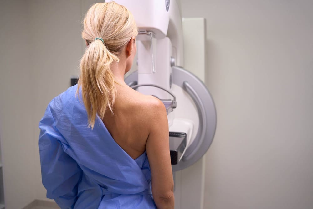
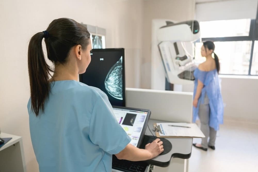
Digital Breast Tomosynthesis (3D Mammography) takes multiple low-dose X-ray images from different angles to build a three-dimensional picture of the breast, which is more accurate than standard 2D mammography at detecting small tumours.
Breast Ultrasound uses high-frequency sound waves rather than radiation to image breast tissue. It is particularly effective at distinguishing solid masses from fluid-filled cysts, and at picking up abnormalities that might blend into dense tissue on a mammogram.
Used together, the two methods complement each other's blind spots: mammography is excellent at detecting calcifications, while ultrasound is better at characterising masses within dense tissue. This is why the combination, rather than either test alone, is recommended for women with dense breasts.
For women without dense breast tissue, FV Hospital's Standard Breast Cancer Screening with 3D mammography alone is typically sufficient.
You check in at FV Hospital's Breast Care Centre. The 3D mammography is performed first, followed by the breast ultrasound, together taking approximately 30 to 40 minutes. A Breast Specialist then reviews both sets of images and discusses findings with you in English. The entire visit is private and conducted in a comfortable, supportive setting, with interpretation available on request in French, Japanese, Korean, and Chinese.
The following add-on can be booked alongside this screening (price converted from FV's VND pricing at approximately 27,000 VND per USD, rounded up):
The add-on price above is converted from Vietnamese dong at approximately 27,000 VND per USD and rounded up to the nearest dollar; the exact amount charged follows FV Hospital's official VND pricing at the time of your visit. All prices are per person and subject to change without prior notice. For non-menopausal women, the best time to perform mammography is the second week of your menstrual cycle.
FV Hospital was established in Ho Chi Minh City in 2003 and is a member of Thomson Medical, one of Southeast Asia's leading healthcare groups. FV was the first hospital in southern Vietnam to earn the JCI Gold Seal in 2016, and has renewed its JCI accreditation four consecutive times with over 98 percent compliance across more than 1,200 international patient safety and quality criteria.
With 45+ specialties, 210+ Vietnamese and international doctors, and care for more than 6 million patients since 2003, FV blends French and Vietnamese medical expertise and provides English- and French-speaking staff for international patients.